

REGION SCANNED:
Extended field both jaws
REASONS FOR REFERRAL:
Implant treatment proposed in both jaws. Bone and maxillary sinus assessment.
SCANNER USED:
KaVo OP 3D V17
SCANNING PROTOCOL:
16×10 cm FOV, 0.25 mm voxel size, 120 kV, 5 mA, 7.4 seconds
RADIATION DOSE:
788 mGy cm2 (approximately 0.15 mSv)
View the full scan using the links below:
View on our PACS web viewer
i-CAT Vision (study name: caseoftheweek19)
RAW DICOM Files
PDF (Maxilla)
PDF (Mandible)
invivo6 (free viewer .exe file)
Findings:
The UR8-6 region has severe loss in vertical alveolar ridge height. The UR5 has widening of the apical periodontal ligament space with evidence of apical pathology. This could be a fibrous scar, an apical granuloma or radicular cyst secondary to chronic apical periodontitis.
The LL7-6 region has moderate loss in vertical alveolar ridge height.
The LR7 region has moderate loss in vertical alveolar ridge height.
Severe periodontal disease is affecting the lower anterior teeth, UL6 and UL7 with loss of attachment of the latter.
The right maxillary sinus has severe mucosal thickening with erosion of the sinus cortical floor and part of the alveolar ridge in the UR6 region. ENT referral is suggested to rule out malignancy.
Both TMJs have severe degenerative joint disease with condylar osteophytes and flattening of the right glenoid fossa. The left condylar head has an Ely’s cyst (also called a subcortical or subchondral cyst).
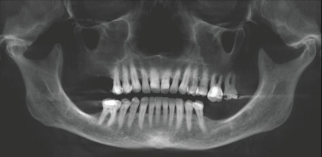
CBCT reconstructed panoramic image.
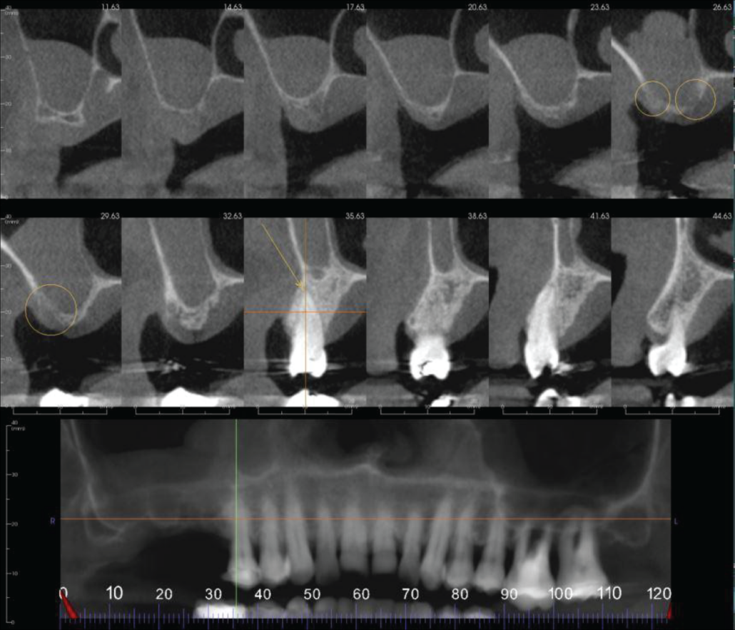
Cropped panoramic and cross sections of the UR8-4 region. The UR8-6 has severe loss in vertical alveolar ridge height. The UR6 region has erosion of the maxillary sinus cortical floor and alveolar ridge (circled). The UR5 has widening of the apical periodontal ligament space (arrow).
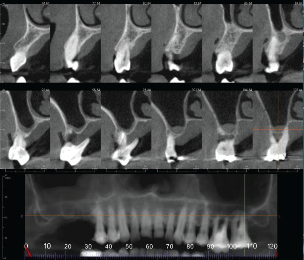
Cropped panoramic and cross sections of the UL6-7 region. The UL6 and UL7 have severe loss of alveolar bone due to periodontal disease.
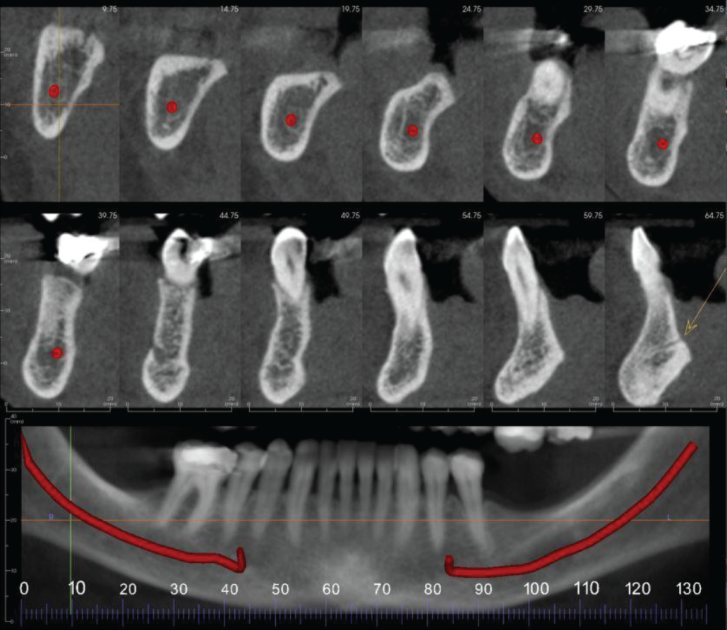
Cropped panoramic and cross sections of the LR8-1 region. The LR7 region has moderate loss in vertical alveolar ridge height. The midline lingual vascular canal is identified by an arrow.
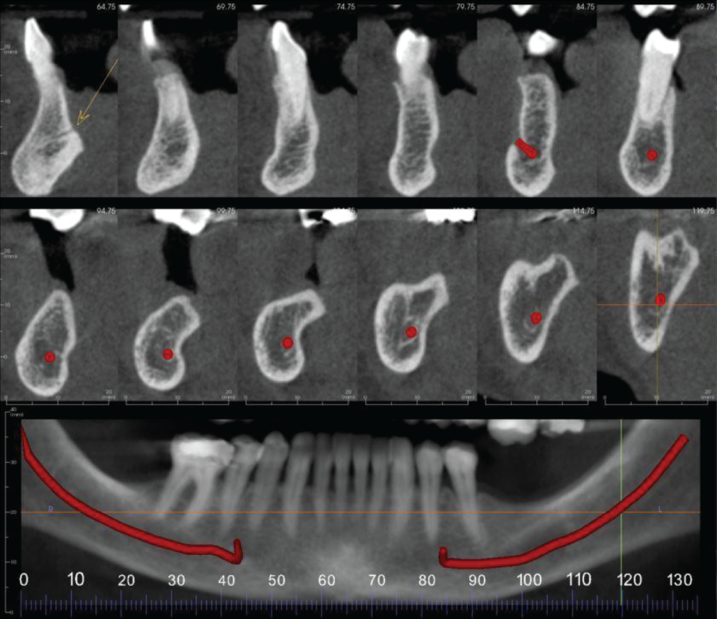
Cropped panoramic and cross sections of the LL1-8 region. The LL6-7 region has moderate loss in vertical alveolar ridge height. The midline lingual vascular canal is identified by an arrow.
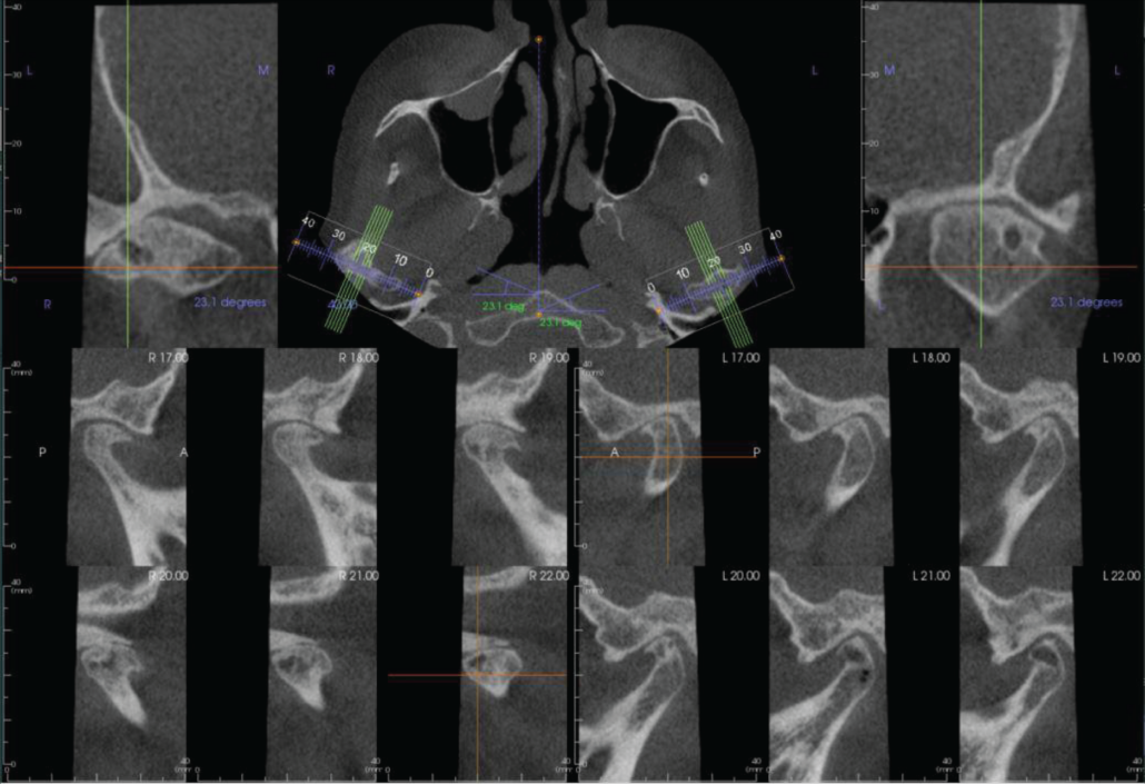
Bilateral parasagittal sections of the TMJs. Both joints have severe degenerative joint disease with condylar osteophytes and flattening of the right glenoid fossa. The left condylar head has an Ely’s cyst.
|
View our other cases 👉 Cases of the week │ Contact Us │ Locations │ Online Bookings

