

REGION SCANNED:
Anterior maxilla UR4 to UL5
REASONS FOR REFERRAL:
Assessment of UL2 before root canal re-treatment
SCANNER USED:
KaVo OP 3D
SCANNING PROTOCOL:
5×5 FOV, 0.125mm voxel size, 95kV, 3.2mA, 9.9seconds
RADIATION DOSE:
304mGy cm 2 (approximately 0.04m Sv)
View the full scan using the links below:
View on our PACS viewer
i-CAT Vision (study name:caseoftheweek20)
RAW DICOM Files
PDF
invivo6 (free viewer .exe file)
Findings:
The UL2 is root-filled. The filling material extends to the apex, with extrusion of a strand of sealer or a very thin gutta percha point for 4mm beyond the apex into a well-defined ovoid radiolucency, about 10mm in diameter. The lesion extends to the palatal and labial cortices, which are both thinned and probably perforated in places. There is slight expansion of the labial cortex (image 2).
The lesion extends mesially to involve the UL1 root apex and reaches the nasopalatine canal. The upper margin of the lesion is clear of the nasal floor (image 3).
A large terminal branch of the anterior superior dentoalveolar canal runs about 2mm palatal to the lesion along the maxillary antral anterior wall (image 4).
The appearance of the lesion is suggestive of either a large periapical granuloma or a small radicular cyst, with secondary involvement of the UL1 root and has created a bone defect across the alveolus.
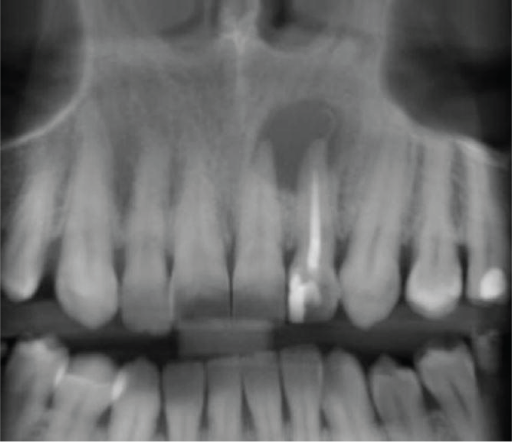
CBCT reconstructed panoramic image
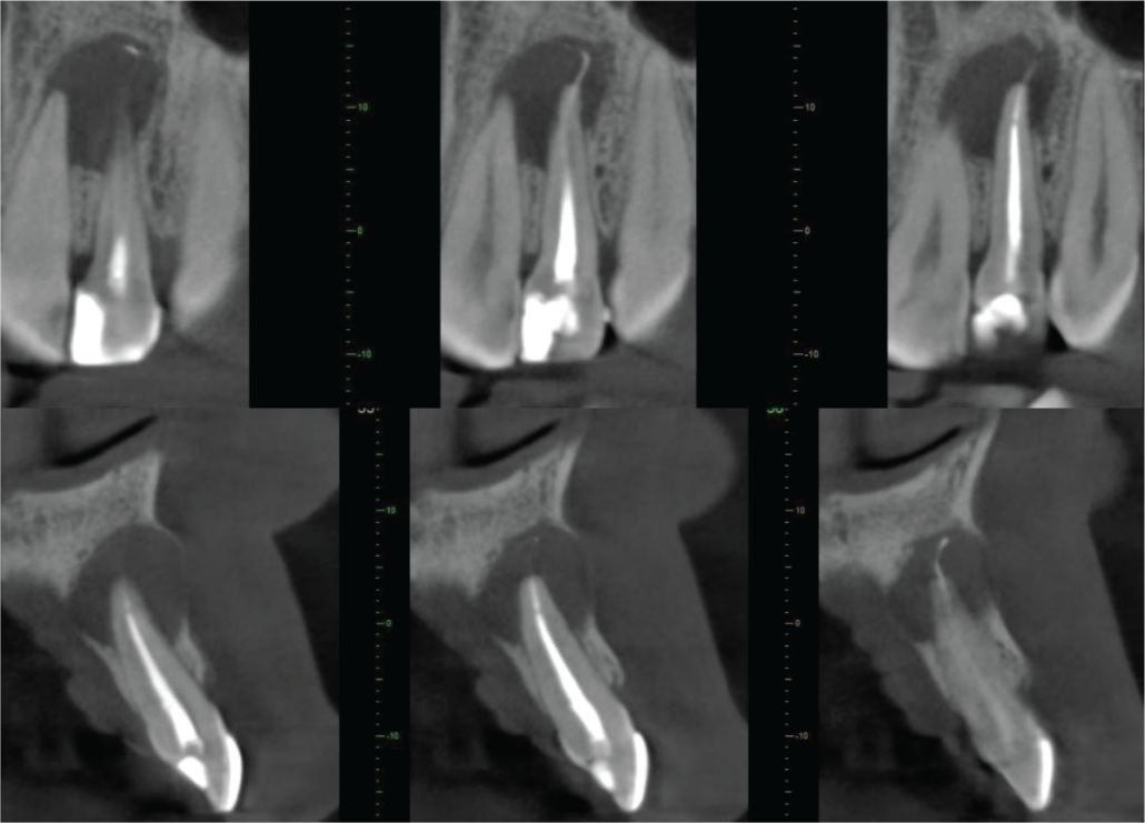
Cross-sections of UL2
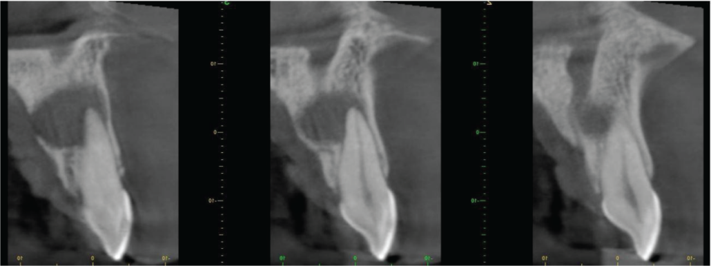
Labio-palatal cross-sections of UL1
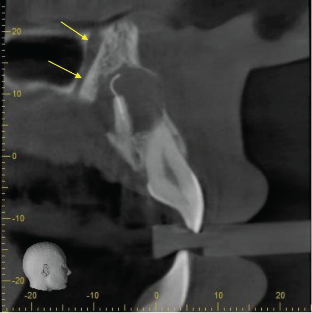
Labio-palatal cross-section to show the prominent neurovascular canal
|
View our other cases 👉 Cases of the week │ Contact Us │ Locations │ Online Bookings

