

Region scanned:
UL1 to UL7
Reason for referral:
Assessment of UL4 implant
Scanner used:
KaVo OP 3D
5x5cm FOV, 0.2mm voxel size, 95kV, 9mA, 2.8seconds
Radiation dose:
267mGy cm2 (approximately 0.03mSv)
View the full scan using the links below:
- View on PACS Browser Viewer
- i-CAT Vision (Study Name:CaseOfTheWeek21)
- Raw Multiple DICOM files
- Raw Single DICOM file
- Xelis Dental Viewer (Version of “On Demand” with FULL Implant Library)
- Invivo6 (Free Viewer)
Findings:
The implant shows definitive signs of failure. There are mesial, palatal and distal vertical bone defects over the coronal half of the implant and a thin radiolucent space around the apical half of the implant on those surfaces. The buccal side of the implant appears denuded of bone, although it is possible that there is some thin bone that the scan is not revealing. There is no point at which osseointegration can be inferred (images 2, 3 and 4).
There is generalised moderate periodontal bone loss with a horizontal pattern.
The UL2 has a flat root end, consistent with apical resorption. There is no associated bony pathosis, so this is probably old and non-progressive resorption (image 5).
There is a mild inflammatory mucosal thickening or a mucous retention cyst on the left antral floor above the molars.
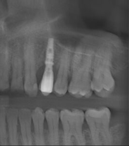
Panoramic reconstruction
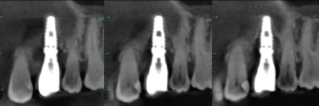
Image 2. Mesio-distal cross sections of UL4 implant
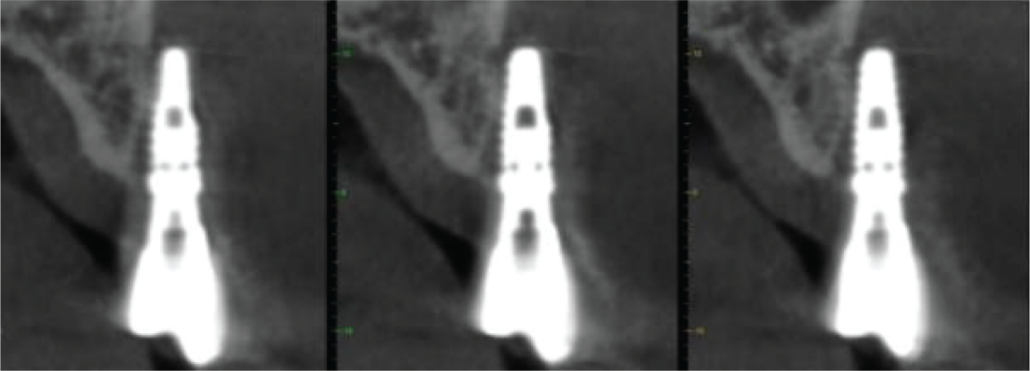
Image 3. Bucco-palatal cross sections of UL4 implant
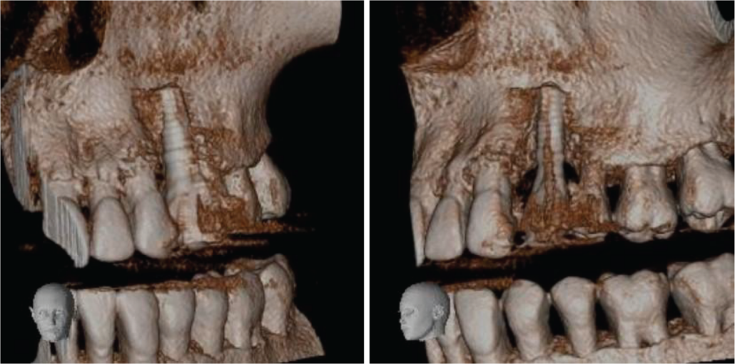
Image 4. Volume-rendered images of UL4 region
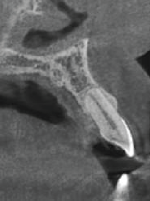 Image 5. Bucco-palatal cross section of UL2
|

View our other cases 👉 Cases of the week │ Contact Us │ Locations │ Online Bookings

