

Region scanned:
UR8 to UL8
Reason for referral:
Investigation of cystic lesion in upper right premolar area
Scanner used:
KaVo OP 3D Vision V17
8x5cm FOV, 0.125mm voxel size, 120kV, 5mA, 7.4 seconds
Radiation dose:
341 mGycm2 (approximately 0.05mSv)
TRY OUR DIFFERENT FORMATS TO LOOK AT THIS CASE:
i-CAT Vision (workup: CaseOfTheWeek22)
*NEW!!! Xelis Dental (full implant library)
Findings:
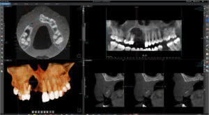
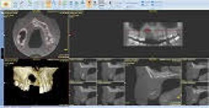
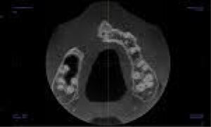
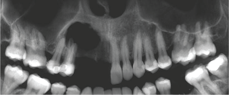
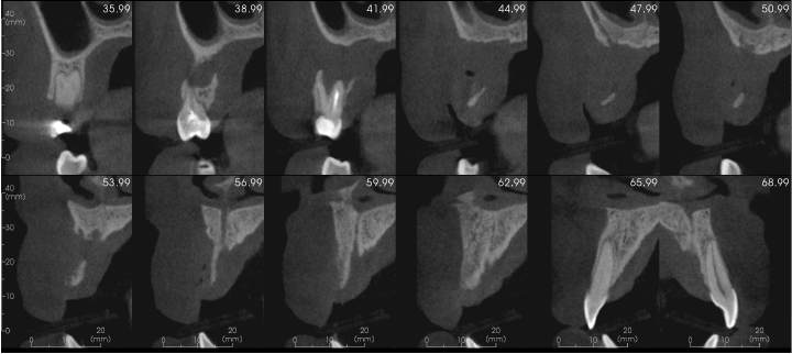
There is a large unilocular moderately well-defined radiolucency, 23 x 15 x 14 mm extending from the UR5 apex to the midline but not invading the nasopalatine canal. There is a “through and through” destruction of the buccal and palatal cortical bony plates, as well as destruction of the alveolar bone ridge. The right maxillary sinus and floor of the nose are not involved. There is external root resorption of the UR4.
The lack of bone expansion, resorption of UR4 roots plus the “floating tooth” appearance of UR4 all suggest the possibility of malignancy. In a case such as this an urgent referral to an oral surgeon is recommended for biopsy to determine the diagnosis and rule out a malignancy.
Panoramic reconstruction
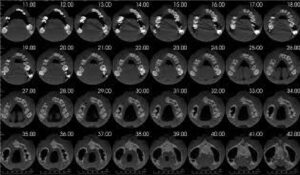
Bucco-palatal cross sections with 3mm spacing between UR1 and UR5
|

View our other cases 👉 Cases of the week │ Contact Us │ Locations │ Online Bookings

