

Region scanned:
UR4 to UL6
Reason for referral:
Assessment of UL2, which appears to have an additional canal
Scanner used:
DEXIS OP 3D
5x5cm FOV, 0.125mm voxel size, 95kV, 3.2mA, 10 seconds
Radiation dose:
304mGy cm2 (approximately 0.04mSv)
Findings:
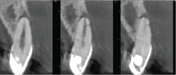
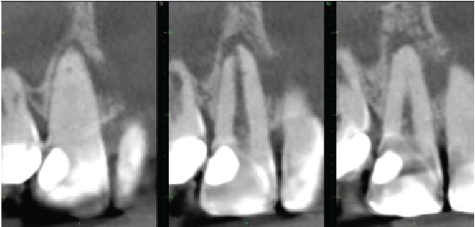
The UL2 has a heavily restored crown. Dens invaginatus anomaly seen (Oehler type I). The root canal is patent. Projections of the root canal pass mesially and distally around the deep part of the invagination and presumably meet on the palatal side. There is also a lateral canal 2.5mm from the apex directed mesiolabially. There is a 7.5mm maximum diameter periapical radiolucency, extending along the mesial side of the root almost to the crestal bone level. The apex is flattened, indicating some apical inflammatory resorption.
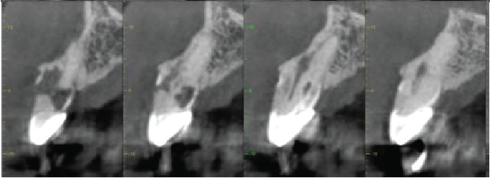
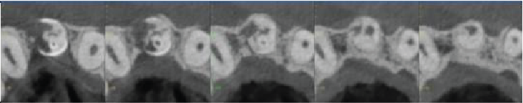
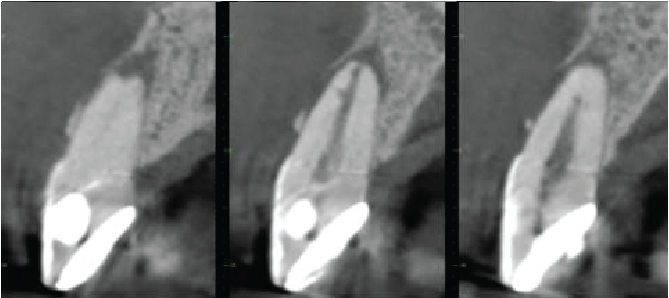
The UR2 is incompletely erupted, with its cervical margin more superiorly placed than on adjacent teeth, with the periodontal bone level correspondingly placed. The tooth is crowned. Dens invaginatus anomaly also seen (Oehler type I), matching that on the contralateral tooth. There is extensive external cervical root resorption arising distally, with a large surface defect and with root canal involvement. The difficulty in seeing the periodontal ligament around the apical half of the root probably indicates some areas of ankylosis. No periapical inflammatory pathosis.
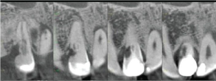
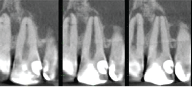
The UR1 is heavily restored. The root canal is patent, with a lateral canal directed mesiolabially 3mm from the root apex. A periapical granuloma is extending down the mesial and labial sides of the root, with a fenestration of the labial cortical plate. There is apical inflammatory root resorption.
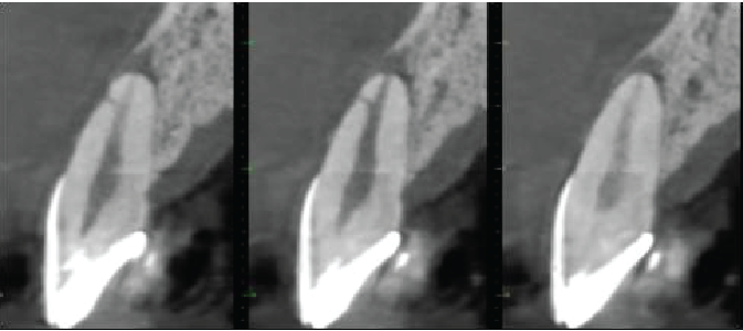
The UL1 has a very similar appearance to the UR1. The root canal is patent, with a lateral canal directed labially 2mm from the root apex. A periapical granuloma is extending down the labial side of the root, with a large fenestration of the labial cortical plate. There is apical inflammatory root resorption.
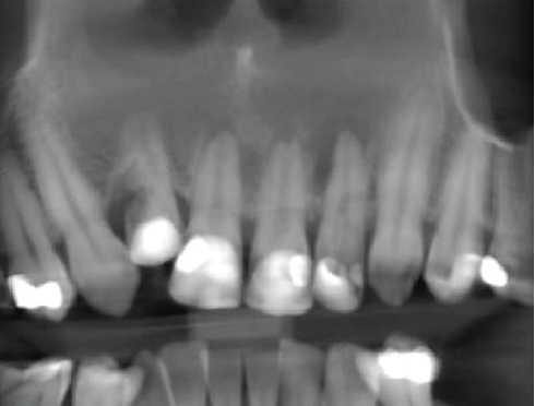
Panoramic reconstruction
Cross sectional images of UL2
Mesio-distal
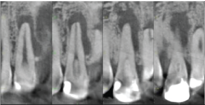
Labio-palatal
Oblique (mesio-labial to distopalatal) to show lateral canal
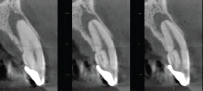
Cross sectional images of UR2
Mesio-distal
Labio-palatal
Axial
Cross sectional images of UR1
Mesio-distal
Labio-palatal
Cross sectional images of UL1
Mesio-distal
Labio-palatal
|

View our other cases 👉 Cases of the week │ Contact Us │ Locations │ Online Bookings

