

Region scanned:
UR8 to UL8
Reason for referral:
Assessment of maxilla before implant treatment
Scanner used:
CS 8100 3D
8×5 cm FOV, 0.15 mm voxel size, 90 kVp, 3 mA, 15 seconds
Radiation dose:
Approximately 0.07 mSv
View the full scan using the links below:
i-CAT Vision  (caseoftheweek23)
*NEW!!! Xelis Dental 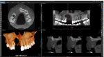 (full implant library)

Findings:
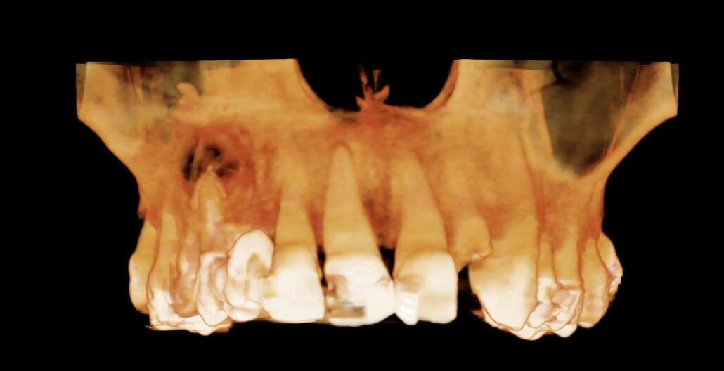
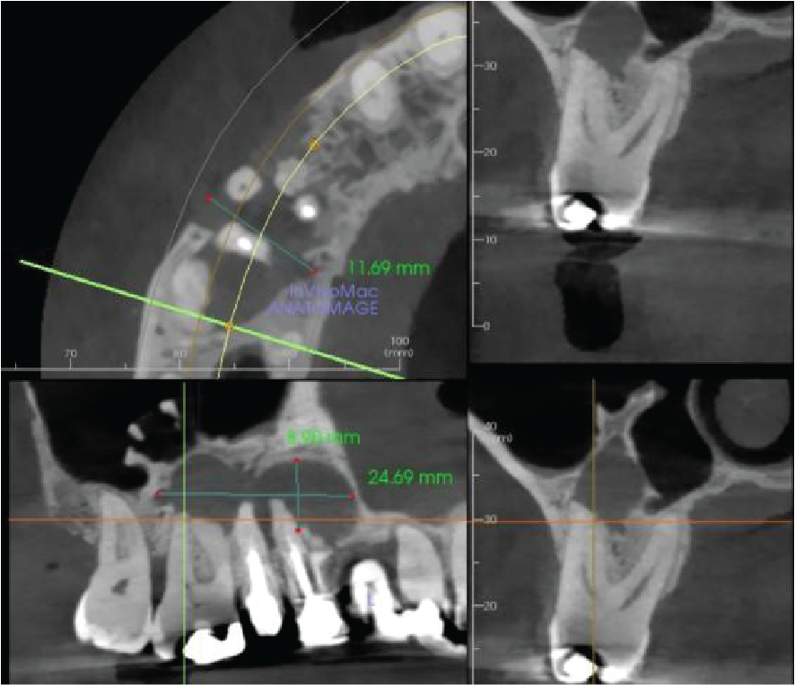
There is apical pathology on the UR6,5,4. The UR6 buccal roots have a large apical radiolucency. The UR4 and UR5 are root treated with large apical radiolucencies and destruction of the buccal cortical plate. These are probably apical granulomas or radicular cysts secondary to chronic apical periodontitis. They are contiguous and measure 12 x 25 x 9 mm.
The right maxillary sinus floor has reactionary bone formation, almost certainly due to the chronic inflammation from the apical disease. However, there is no thickening of the sinus mucosa.
The remaining teeth have mild to moderate generalized alveolar bone loss due to stage II periodontal disease.
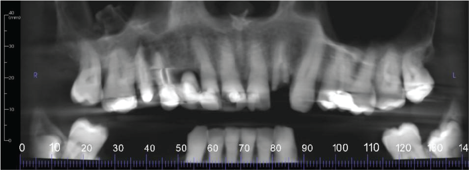
Panoramic Reconstruction
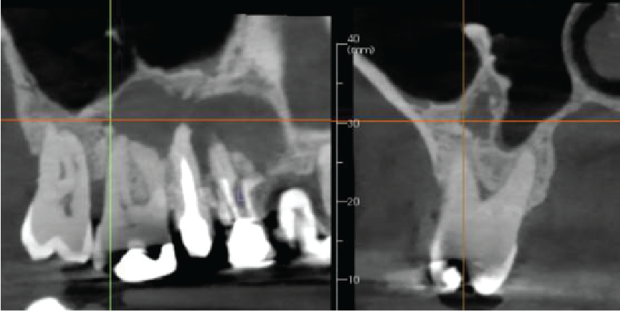
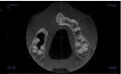
Cropped panoramic and cross section of the alveolar ridge in the UR4-6 region showing chronic apical infection and reactionary bone formation in the right maxillary sinus
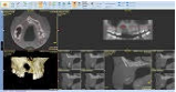
Cropped panoramic, axial and cross sections of the alveolar ridge UR4 6 region showing chronic apical infection
|

View our other cases 👉 Cases of the week │ Contact Us │ Locations │ Online Bookings

