

REASON FOR REFERRAL: Unerupted supernumerary tooth seen on OPG. Assessment with CBCT to check position and impact with planned orthodontic treatment
REGION SCANNED: Mandible, extending from the anterior right ramus to the left first molar region
SCANNING PROTOCOL: 5×9 cm FOV, 0.2 mm voxel size, 95 kV, 4 mA, 20 seconds
RADIATION DOSE: 704 mGycm2
View the full scan using the links below:
Please click here to view on our PACS
Please click here to download i-CAT Vision (Patient name: Caseoftheweek18)
Please click here to download RAW DICOM
Please click here to view as PDF
Please click here to download invivo6
FINDINGS:
There is an unerupted supernumerary tooth lying in a mesioangular position, and with a lingual inclination, in the right premolar region of the mandible (image 2). It is 19.5mm in length.
The crown of the supernumerary tooth is lingual to the middle thirds of the roots of LR4 (44) and LR5 (45), below a thinned lingual cortical plate (image 3). The pericoronal follicle space is of normal size.
The crown of the supernumerary tooth is impacted against the middle third of the root of LR4 (44) (image 4a), while both the crown and root are in contact with the middle and apical thirds of the root of LR5 (45) (image 4b). There is no root resorption seen on LR4 (44) or LR5 (45).
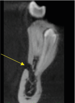
From a surgical perspective, the root of the supernumerary tooth is straight and its apex contacts the ID canal at the point at which the mental canal is given off (images 5 and 6). The only potential risk would be with downward pressure. In a surgical context there is a potential risk with any applied downward pressure.
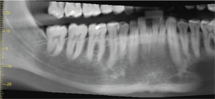 1. Panoramic reconstruction. The supernumerary tooth is barely visible due to its lingual position.
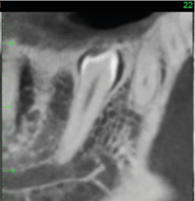
2. The supernumerary tooth on mesio-lingual cross-section.
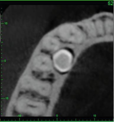
3. Axial cross-section showing the crown of the supernumerary tooth (image viewed from below).
4. Mesio-distal cross-section through LR4 and LR5 roots, showing the crown of the supernumerary lingually.
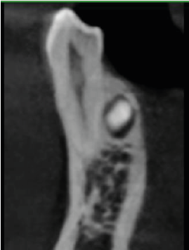
a. Through LR4 root, with part of the crown of the supernumerary lingually
a. Through LR4 root, with part of the crown of the supernumerary lingually
5. Oblique mesio-distal cross-section through the supernumerary tooth, showing the neurovascular canal in contact with its root apex (mental foramen arrowed).
6. Volume-rendered image of buccal surface of mandible showing mental foramen position and two Maximum Intensity Projection images with the ID and mental canals marked.
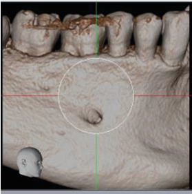
Volume-rendered image
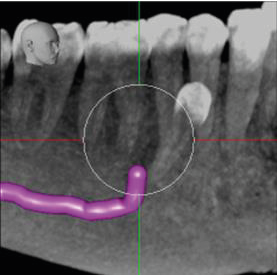
MIP image from the buccal perspective
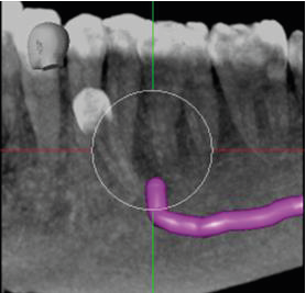
MIP image from the lingual perspective

