
![]()
Region scanned:
UR4 – UL4
Reason for referral:
Symptomatic root canal filled UR1. Microsurgery planned
Scanning Protocol:
5×5 cm FOV, 0.12 mm voxel size, 95 kV, 4 mA, 10 seconds
Radiation dose:
380 mGycm2 (approximately 0.05 mSv)
View the full scan on your PC:
i-CAT Vision Format (study name: CaseofTheWeek17)
Raw DICOM files
Invivo6 Viewer (.exe file software included)
PDF template
PACS Viewer
Findings:
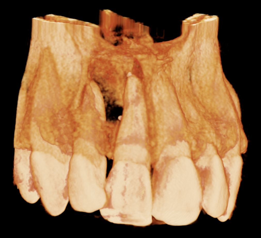
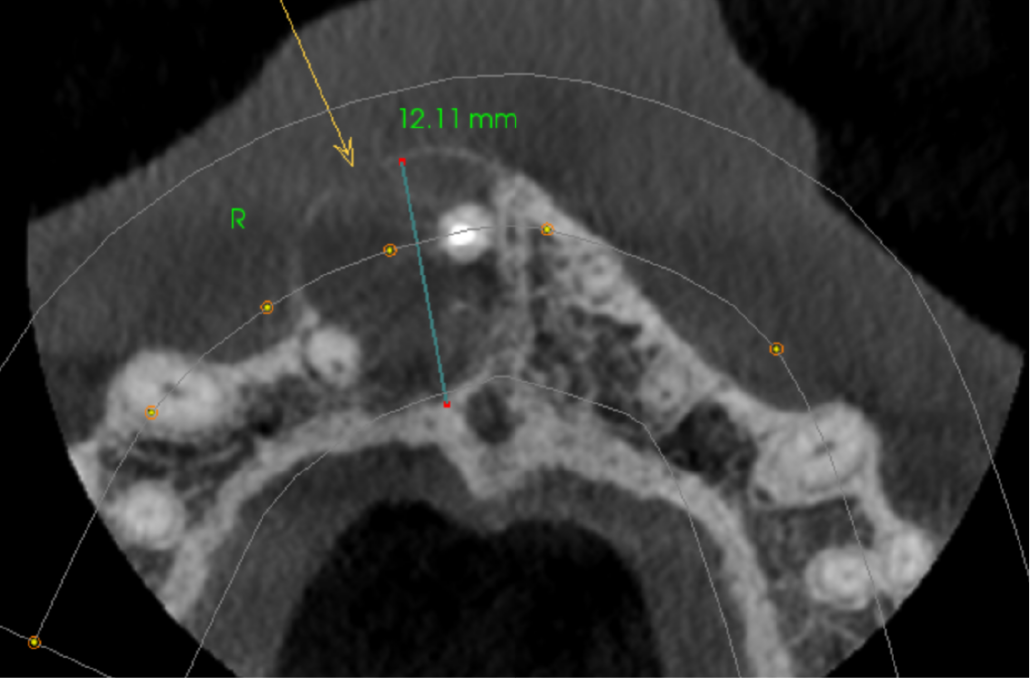
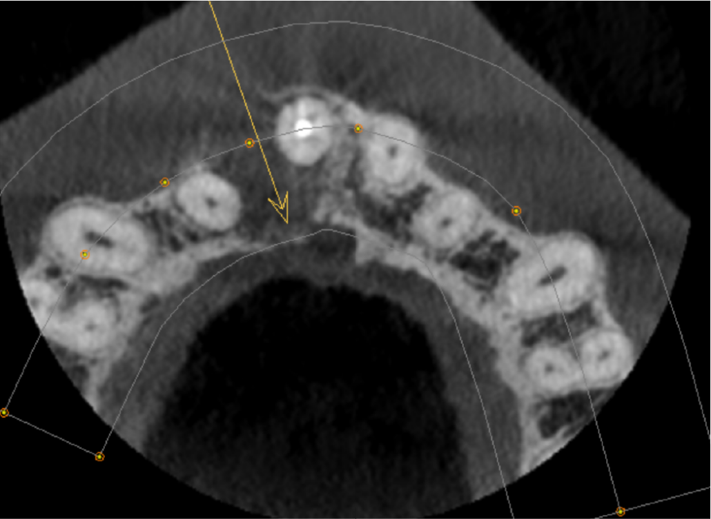
The UR1 is root filled with a small amount of extruded filling material inside the lesion. There is a well-defined, unilocular radiolucency measuring 12mm, extending from the UR1 apical region to the UR2 apex. The origin is probably from the apex of the UL1. The buccal cortex is expanded and perforated. The palatal cortex is eroded at the level of the oral opening of the nasopalatine canal. The lesion has not eroded the nasopalatine canal more superiorly. The appearance is that of a radicular cyst.
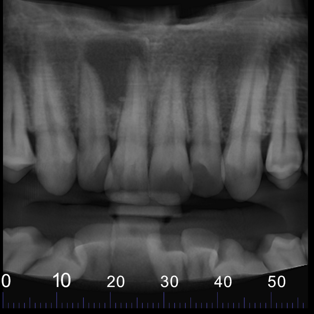
CBCT reconstructed panoramic image
Axial view showing well defined unilocular radiolucency in UR1-UR2 area. The buccal cortex is expanded and perforated (arrow)
Axial view showing the palatal cortex eroded at the level of the oral entrance to the nasopalatine canal (arrow)
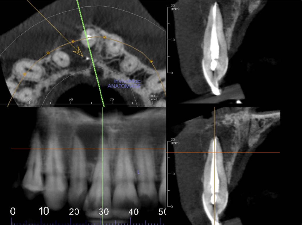
Cropped panoramic, axial and cross section of alveolar ridge UR1 region. UR1 is root filled to the apex with a small amount of extruded filling material inside the lesion (arrow)
|

View our other cases 👉 Cases of the week │ Contact Us │ Locations │ Online Bookings

