

Region imaged:
UL3-UL8
Clinical information:
Periapical pathosis suspected
Scanner used:
KaVo OP 3D
5x5cm FOV, 0.12mm voxel size, 90 kVp, 3.2 mA, 10 seconds
Radiation dose:
Approximately 0.04 mSv
View the full scan using the links below:
i-CAT Vision  (caseoftheweek25)
*NEW!!! Xelis Dental 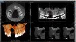 (full implant library)
Findings:
Region UL3-UL8 is imaged. The UL6 is absent. “Pillow”-like mucosal swellings on the floor and walls of the left maxillary sinus, indicating polypoidal mucosal thickening. This appearance is consistent with chronic rhinosinusitis. The apices of the UL7 and UL8 have an intimate relationship with the antral floor and the Schneiderian membrane.
The roots of the UL3/4/5 demonstrate hypercementosis. Image 1
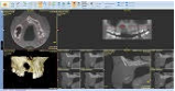
The UL4 has two roots which are practically fused due to hypercementosis. The buccal root is straight. The palatal root has a slight buccal curve. The apical half of both roots is not visible (possibly obliterated). Image 2
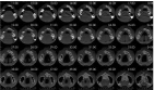
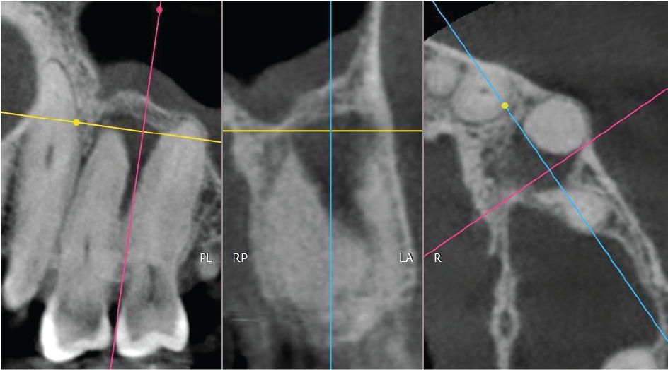
The UL5 has two roots which are practically fused due to hypercementosis. The apical half of the buccal root is not visible (possibly obliterated). Image 3
Between the UL4 and UL5 there is a well-defined radiolucency measuring approximately 6mm diameter. The lesion envelops the UL4 apex and abuts the apical half of the UL5 root. The genesis of the radiolucency is not of obvious endodontic origin since both teeth are unrestored and the location is not typically centred around the apex. If the teeth are vital, suggested tentative diagnosis are odontogenic cyst such as lateral periodontal cyst or fibro-osseous origin. Image 4
Image 1
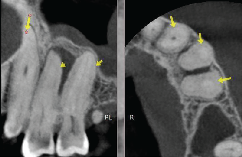
Image 2
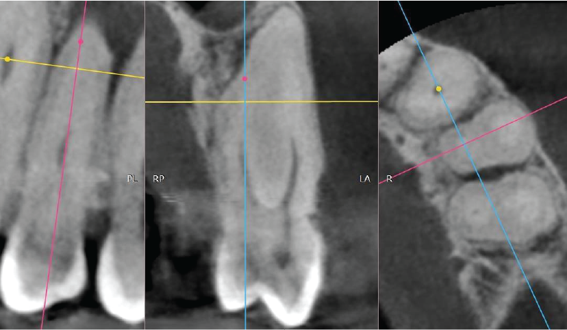
Image 3
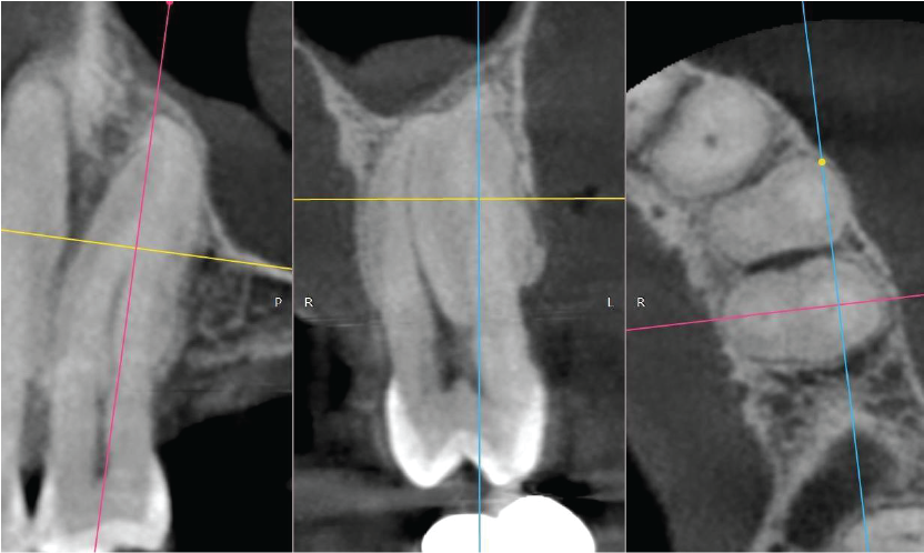
Image 4
|

View our other cases 👉 Cases of the week │ Contact Us │ Locations │ Online Bookings

