

![]()
Xelis Dental Viewer (Same price as i-CAT Vision)
Allows you to plan your patient’s treatment easily, with a full implant library and free virtual implant placement capabilities. Our clinical team will prepare a study for you that includes highlighting the inferior dental canal and image rotation according to the area of interest.
Findings:
The UL1 is displaced buccally with no root fracture. The apical vessels are likely torn due to the displacement. No alveolar fracture is seen, although there is a little buccal cortical plate still attached to the root of UL1.
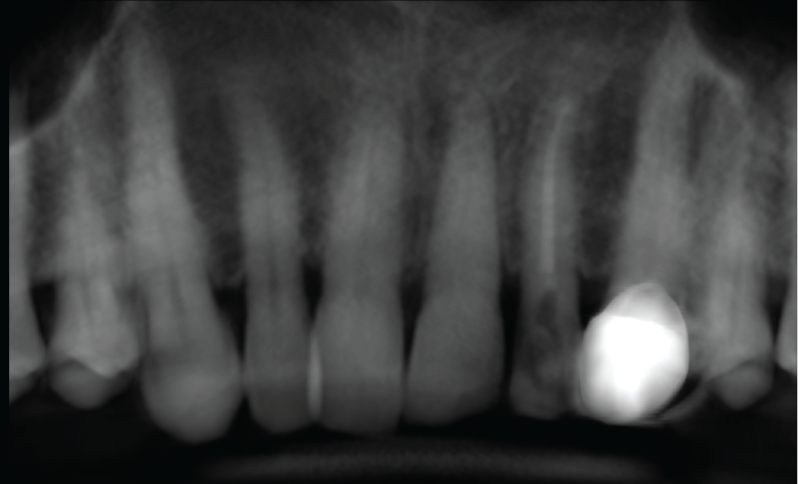
CBCT Reconstructed Panoramic Image
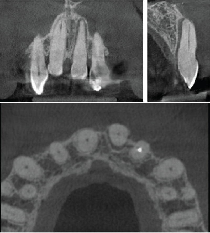
Coronal, sagittal and axial slices through the UL1
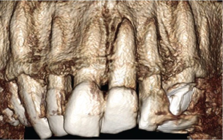
Volume rendered image showing the UL1
Learning from this case:
Small volume CBCT can be used additionally to 2D X-rays in the diagnosis and management of dentoalveolar trauma after a fall, accident or other mechanism of injury. CBCT is able to show luxation, root fractures and alveolar fractures.
Thank you for your continued referrals.
|

View our other cases 👉 Cases of the week │ Contact Us │ Locations │ Online Bookings

