
Region imaged:
UR1 – UL6
Clinical information:
Assessment of UL4 site for implant placement. Extraction was 3 months ago.
Scanner Used:
Gendex DP700
Scanning Protocol:
6×4 cm FOV, 0.2 mm voxel size, 90 kVp, 10 mA, 2 seconds
Radiation dose:
Approximately 0.02 mSv
![]()
Xelis Dental Viewer (Same price as i-CAT Vision)
Allows you to plan your patient’s treatment easily, with a full implant library and free virtual implant placement capabilities. Our clinical team will prepare a study for you that includes highlighting the inferior dental canal and image rotation according to the area of interest.
Findings:
The UL4 region has mild loss in vertical ridge height. The buccal cortical plate is thin or absent. There is Misch 4 bone density (Image 2).
The remaining teeth have mild to moderate generalized alveolar bone loss due to periodontal disease.
The nasopalatine canal has a concave surface and the width is at the upper limit of normal (6mm). This may indicate the development of a nasopalatine cyst. Taking a periapical radiograph in 12 months is recommended to check for any enlargement (Image 3).
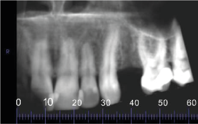
Image 1: Reconstructed Panoramic Image
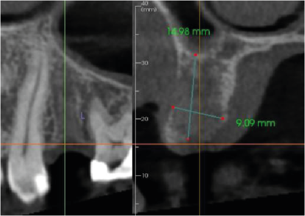
Image 2
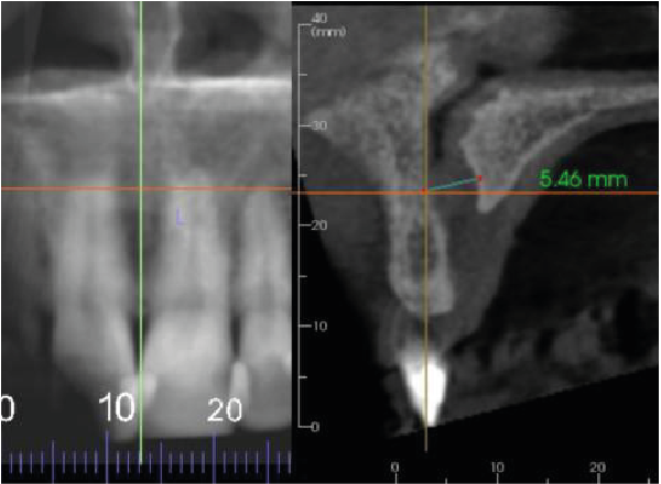
Image 3
Thank you for your continued referrals.
|

 |
 |
 |
 |
 |

