
Scanner Used and Location:
Dental Scan Ltd Manchester Branch
DEXIS OP 3D LX (2023 model) FOV: 5X5cm – 20x15cm
Region Imaged:
LL678
Clinical Information:
Assessment of mesial-buccal defect on LL6
Scanning Protocol:
5x5cm FOV, 0.12mm voxel size, 95 kVp, 5 mA, 11.5 seconds exposure
Radiation Dose:
Approximately 0.07 mSv
![]()
Xelis Dental Viewer (same price as i-CAT Vision)
Allows you to plan your patient’s treatment easily, with a full implant library and free virtual implant placement capabilities. Our clinical team will prepare a study for you that includes highlighting the inferior dental canal and image rotation according to the area of interest.
Findings:
The LL6 mesial-buccal root demonstrates a large root resorption in the cervical third with a wide area of root perforation. There is also another small mesial external resorption.
The LL6 also has periradicular radiolucencies indicating a combined endo-perio lesion. It is most likely unrestorable and has poor prognosis. The inferior dental canal does not contact the LL6 roots.
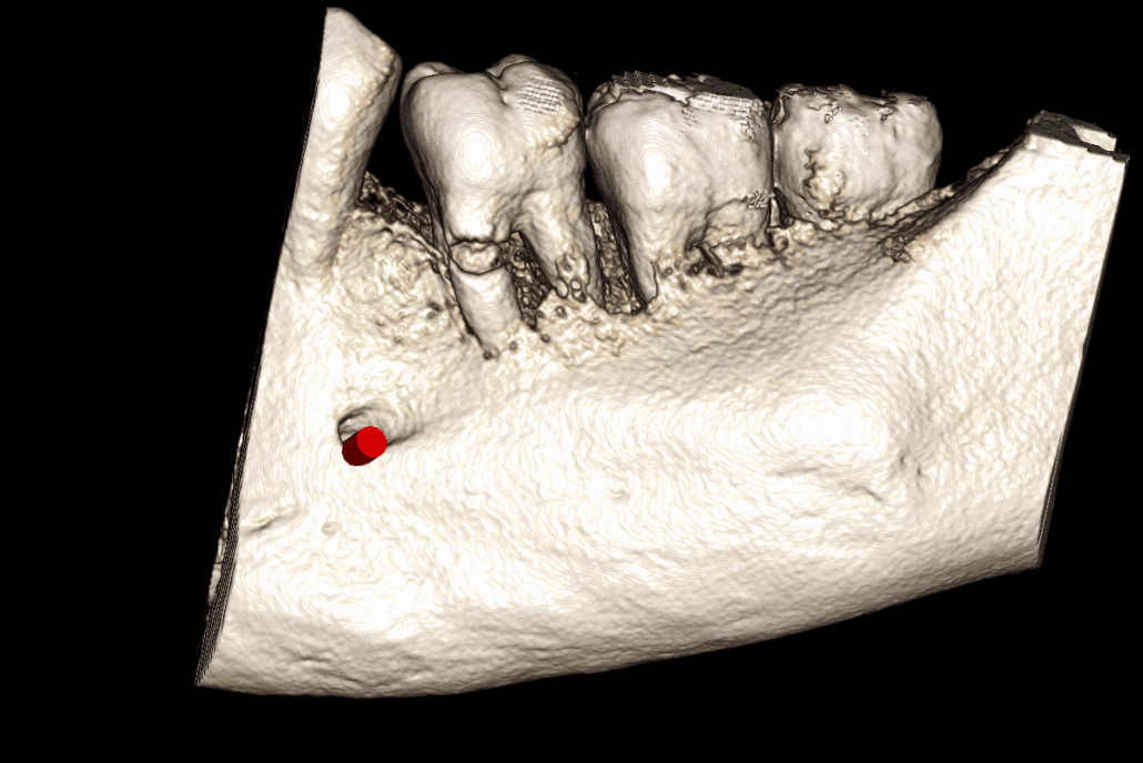
The LL8 roots have a marked distal dilaceration/apical hooks. The upper border of the inferior dental canal contacts the inferior surface of the LL8 roots.
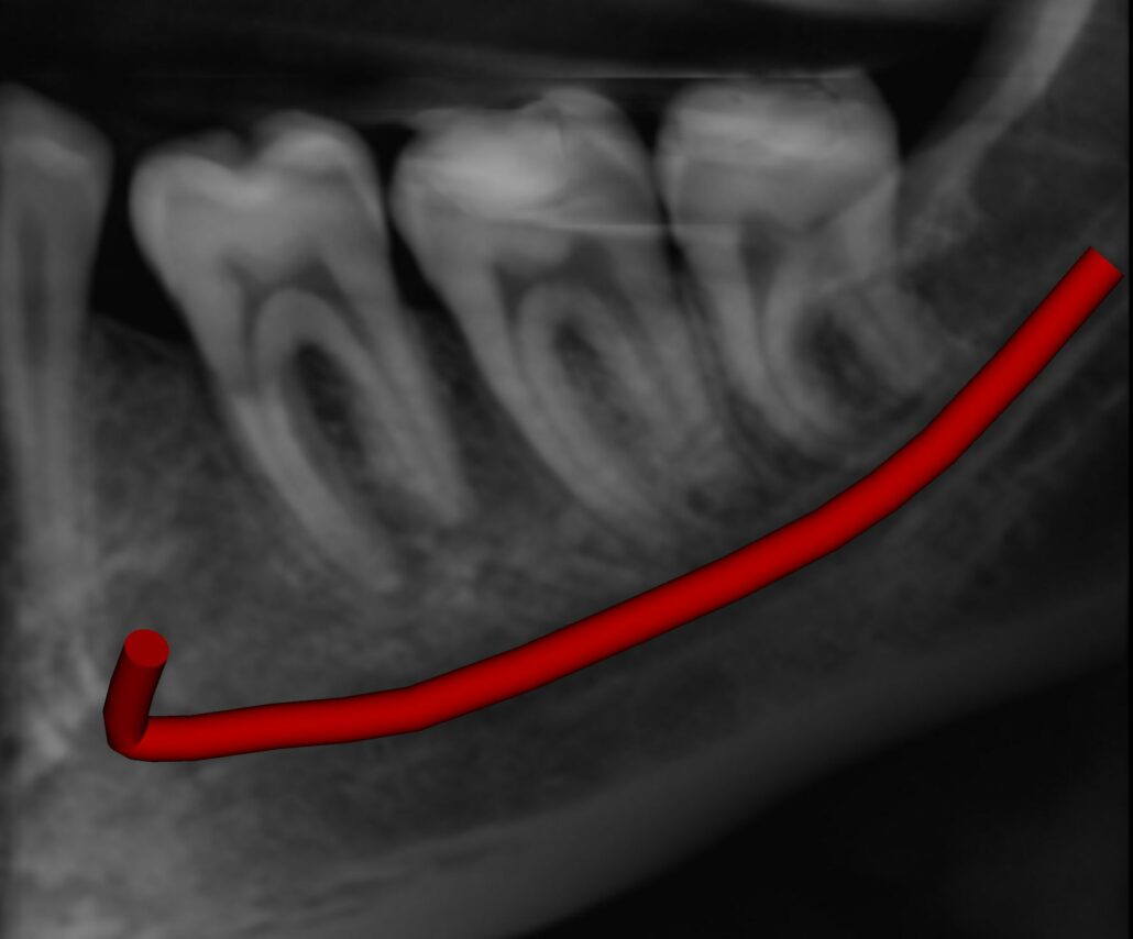
Image 1: Reconstructed Panoramic Image
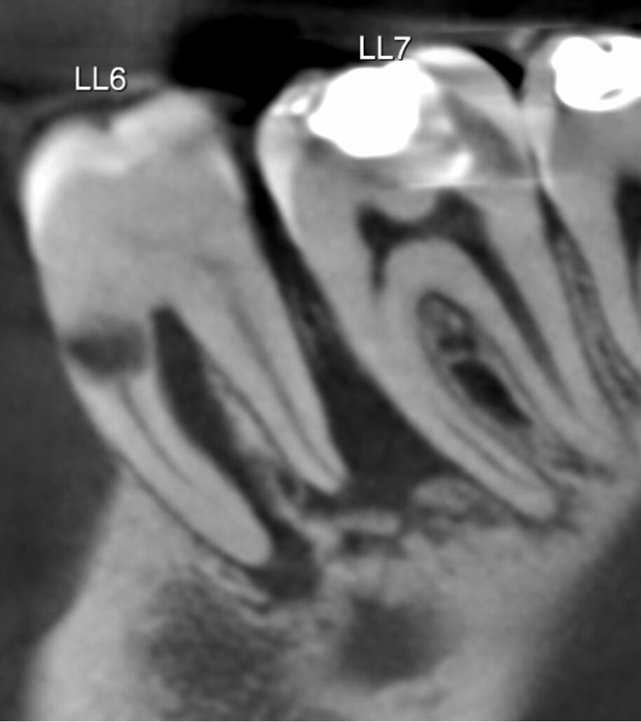
Image 2
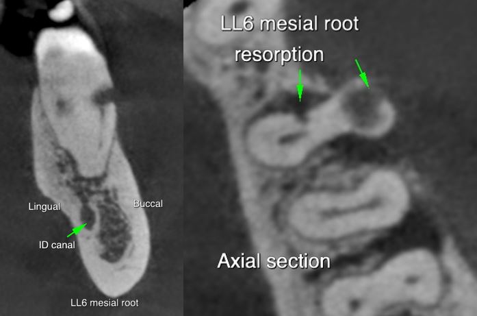
Image 3
Thank you for your continued referrals.
|

 |
 |
 |
 |
 |

