REGION SCANNED: LL3-8
REASON FOR REFERRAL: To assess the relationship of the LL8 with the IDN. OPG inconclusive. Possible infection seen around LL7
SCANNER USED: KaVo OP 3D
SCANNING PROTOCOL: 6.5x9cm FOV, 0.2mm voxel size, 95kV, 3.6mA, 20 seconds
RADIATION DOSE: 896 mGycm2 (approximately 0.13 mSv)
SCAN: Click here to view online
OBSERVATIONS / FINDINGS:
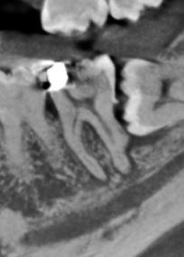
The LL8 is unerupted and horizontally impacted against the LL7 with the mesial root apex touching
the IDC. The tooth has a normal follicle space with no erosion of the adjacent distal root.
The LL7 displays distal calculus and severe loss of periodontal bone attachment. There is mesial horn
pulpal exposure with apical radiolucencies 1-2 mm wide.
There are bilateral mandibular lingual tori.
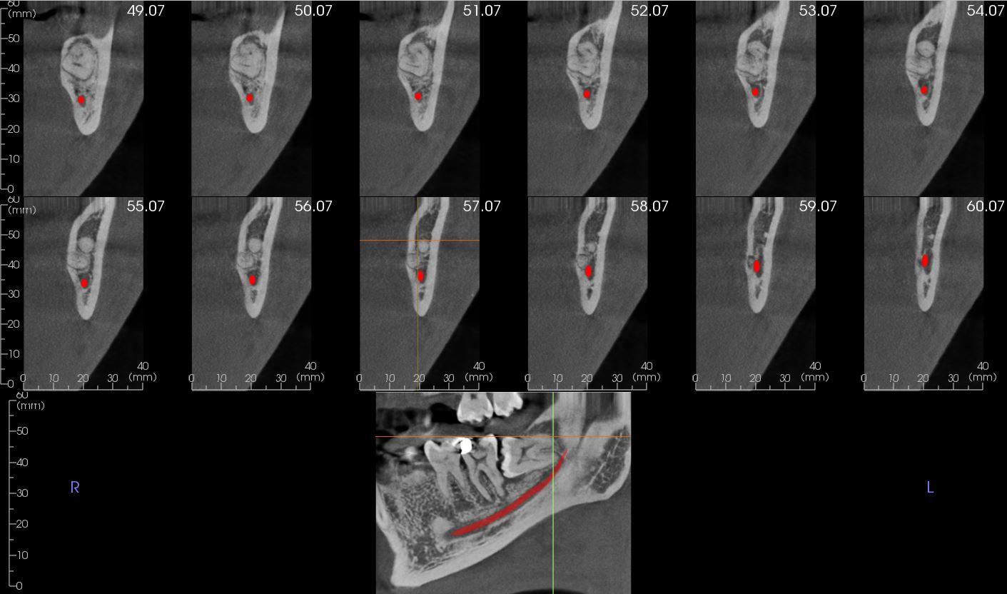
Reconstructed panoramic view and cross sections showing the
relationship of the mesial root apex of the LL8 to the IDN.